Case5 – Persistent NSCLC
Author:
Felix Herth, MD and Ralf Eberhardt, MD, Thoraxklinik, University of Heidelberg, Germany
Source:
DVD-ROM ‘Endoscopic Ultrasound – Diagnostics and Staging of Lung Cancer’, Olympus Europa SE & Co. KG, 2013
Patient History
Male, 68 years. Patient with a history of NSCLC (squamous cell lung cancer) stage II B (T2N1M0) in 2008.
Treatment: surgery and adjuvant chemotherapy. 3 months interval routine follow-up with CT. CT shows enlarged lymph node in station 4R in 2010.
Suspicion of right sided mediastinal tumour.
Treatment: surgery and adjuvant chemotherapy. 3 months interval routine follow-up with CT. CT shows enlarged lymph node in station 4R in 2010.
Suspicion of right sided mediastinal tumour.

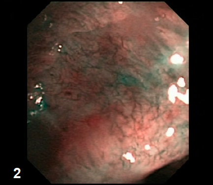
1
RUL stump shows an area with a different colour compared to the surrounding tissue.
2

3
5 mm lesion in the upper area of LN station 4R.
5 mm lesion in the upper area of LN station 4R.
Echopoor lesion of 14×12 mm close to the superior vena cava (LN 4R).
EBUS-TBNA case4
0:55
4
Pathology
Specimen from lymph node station 4R positive for squamous cell carcinoma (NSCLC). Diagnosis
Local lymph node metastasis of persistent NSCLC. Therapy
Radiation therapy.
Specimen from lymph node station 4R positive for squamous cell carcinoma (NSCLC). Diagnosis
Local lymph node metastasis of persistent NSCLC. Therapy
Radiation therapy.
- Content Type