Prof. Ho Khek Yu Lawrence, MD
National University Hospital of Singapore
Professor Lawrence Ho is the Vice Dean of Research and Graduate Studies, Yong Loo Lin School of Medicine, National University of Singapore, and the Group Research Director, Research Office, National University Health System, Singapore. Concurrently, he is a Senior Consultant, Division of Gastroenterology & Hepatology, Department of Medicine, National University Hospital, Singapore, and a Director of the National Referral Laboratories, Pte Ltd. He chairs the Gastroenterology Residency Advisory Committee, and sits in the Joint Commission on Specialist Training, Ministry of Health. He is also president of the Asian EUS Group (AEG).
Overview
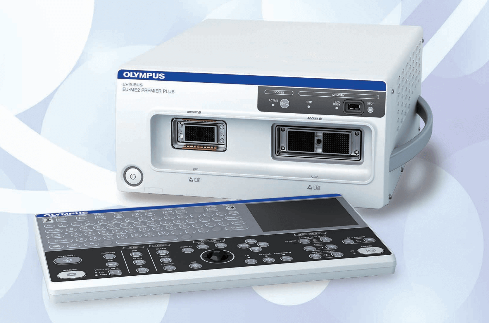
Endoscopic ultrasonography (EUS) is useful for the diagnosis and treatment of pancreatobiliary diseases. It plays a critical role in detecting small lesions (especially those missed by other modalities such as CT), differential diagnosis, tumour staging and various drainage techniques. The Olympus EUS processor (EU-ME2 PREMIER PLUS) has three key functions; namely, tissue harmonic echo (THE), contrast harmonic EUS (CH-EUS) and elastography (ELST), the usefulness of which have been reported.
THE produces high quality images of pancreatic lesions by minimizing the amount of artifacts. CH-EUS when used with a contrast agent, is beneficial for the differential diagnosis of pancreatic tumours. ELST helps to distinguish between benign and malignant lesions by virtue of tissue stiffness.
Despite wide spread awareness of these functions, their actual usefulness is still not well understood. Therefore, this article elucidates the usefulness of THE and ELST with illustrations from actual cases.
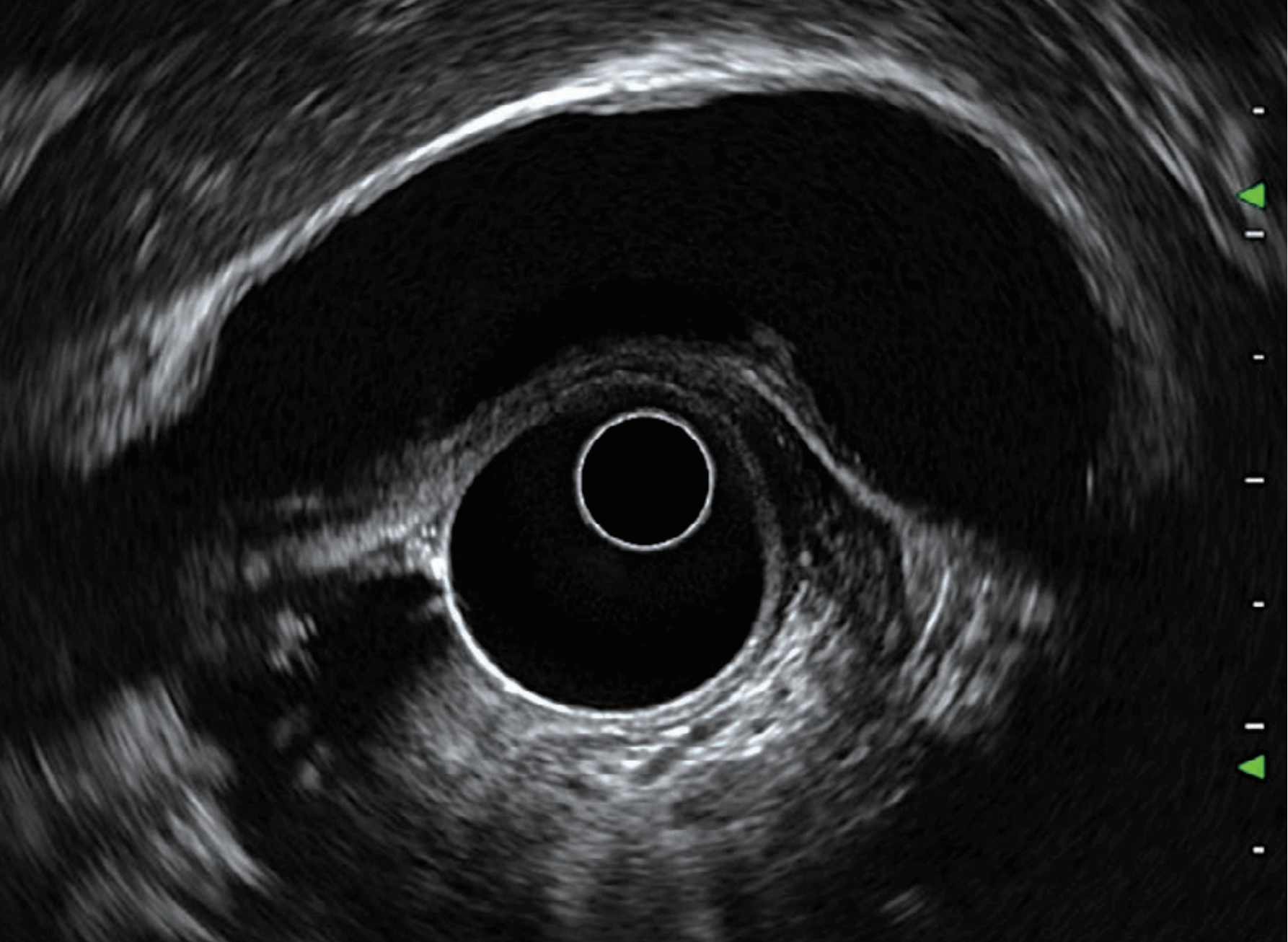
B mode & Elastography (ELST)
Case 1 Pancreatic Adenocarcinoma
A 73-year-old male patient has hypertension, hyperlipidemia and painless obstructive jaundice. CT scan shows a pancreatic head/uncinate mass measuring 3.3 cm x 2.5 cm with biliary tree obstruction which was suspected to be malignant in nature. The mass is seen adjacent to the right hepatic artery. There are several hypodense nodules present in the right liver and caudate, most measuring around 1.4 cm x 1.2 cm and are likely metastases. Endoscopic Ultrasonography (EUS) and Fine Needle Aspirations (FNA) were performed using Olympus’s EU-ME2 PREMIER PLUS. A hypoechoic lesion measuring 2.9 cm x 2.5 cm was observed in the head of pancreas and was seen to be associated with a dilated common bile duct measuring 13 mm. Sludge was present in the distal CBD. The main pancreatic duct was not dilated (2 mm).
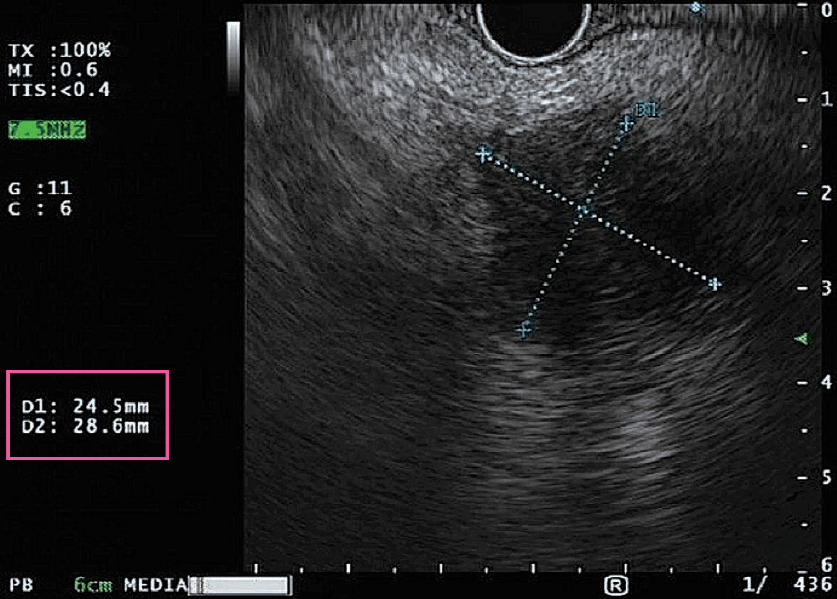

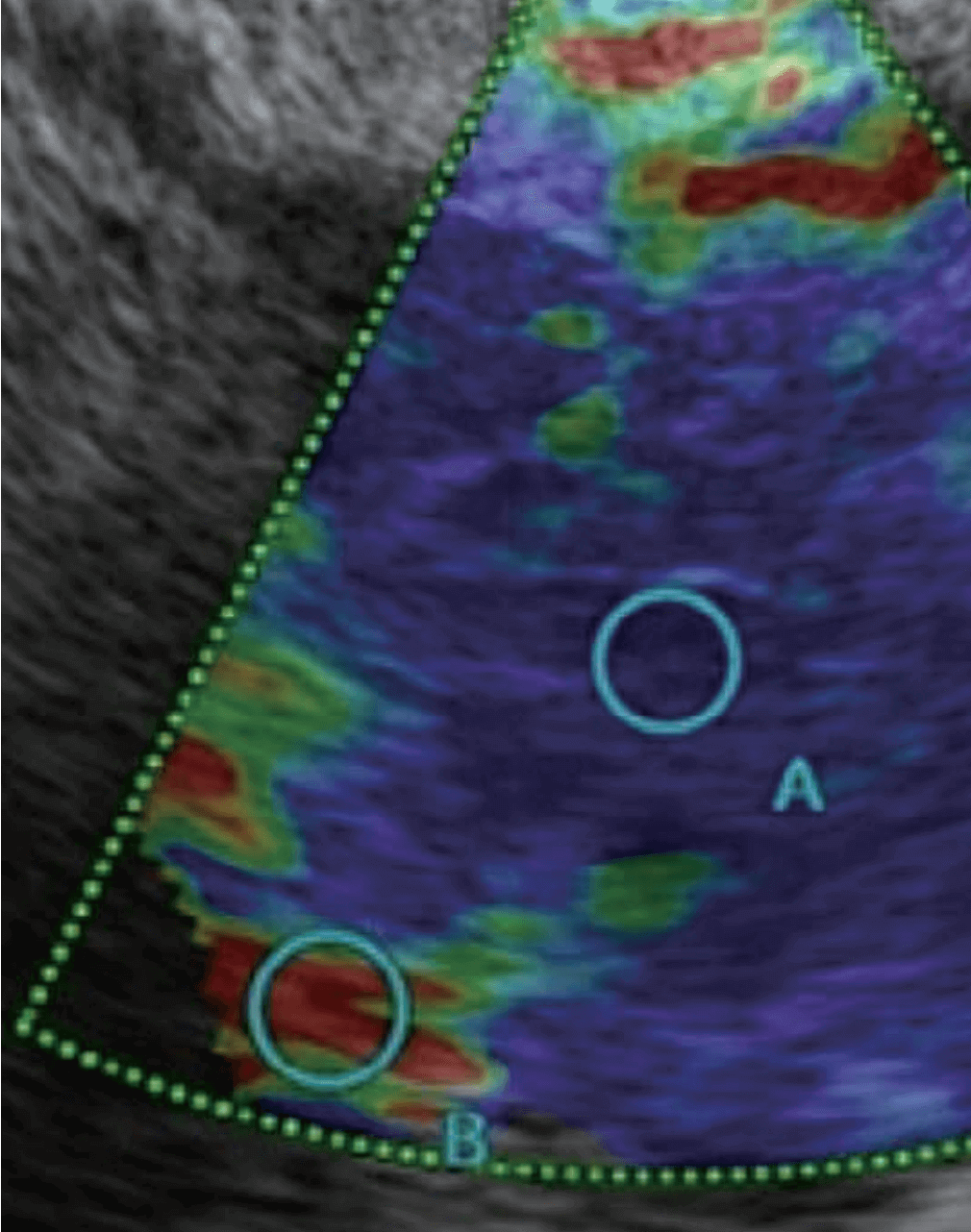
ELST was performed to assess the hardness of the lesion. Area ‘A’ refers to the target pancreatic lesion while area ‘B’ refers to normal tissue. The percentage values of areas “A” and “B” indicate how much they are compressed when the scope tip is being placed against the mucosal surface.
The B/A ratio was measured to be 153.67 which is suggestive of malignancy. To ascertain malignancy, EUS-FNA was performed. 3 passes were done and specimens were sent for cytology. These specimens tested positive for malignant adenocarcinoma.
Therefore, ELST proved useful in identifying the stiffness of the lesion as indicated in blue. This led to highly targeted FNAs which yielded high quality samples. In consideration of better patient care, I recommend the use of ELST prior to performing FNAs for high quality yields.

Technical considerations
B mode
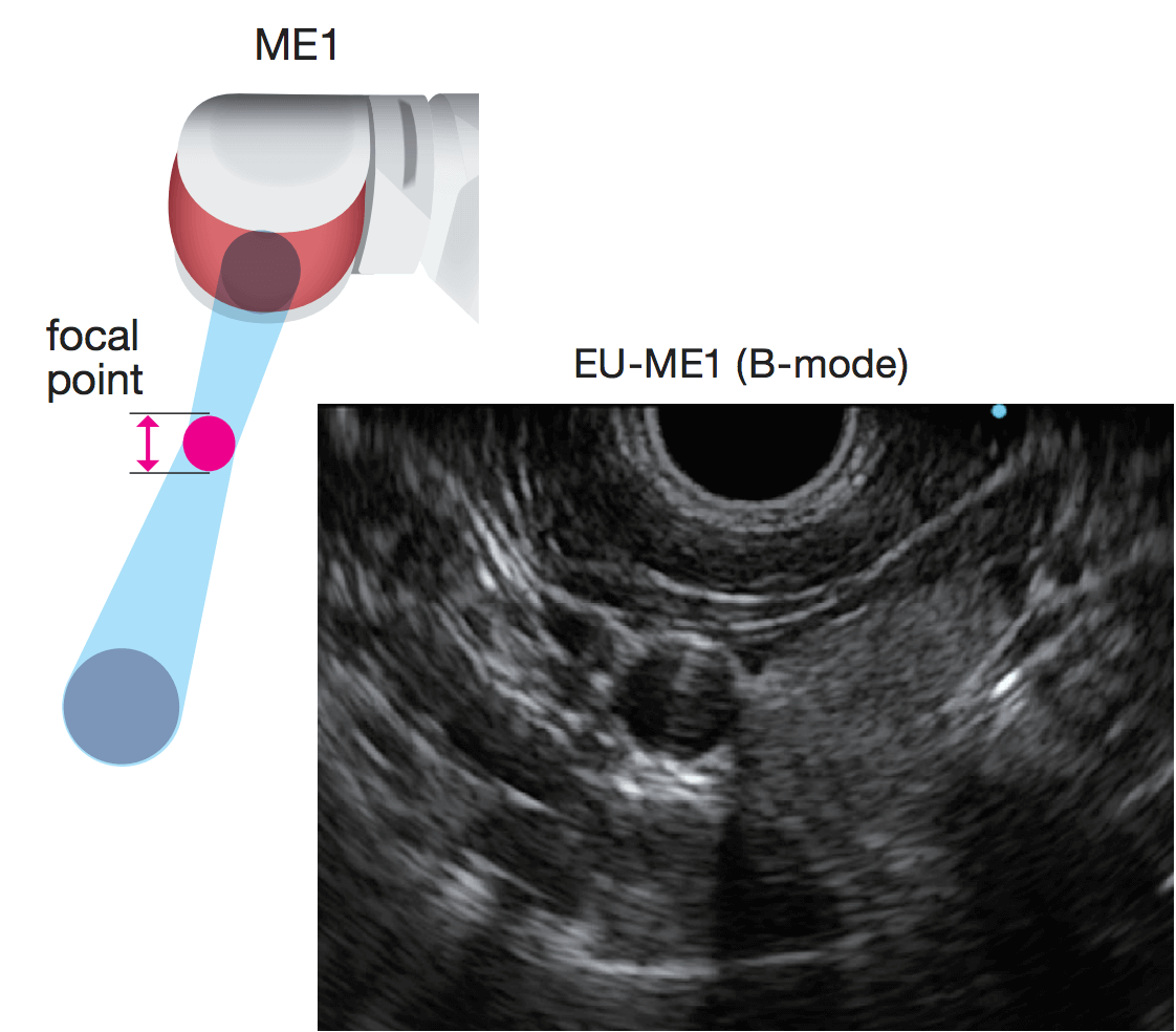
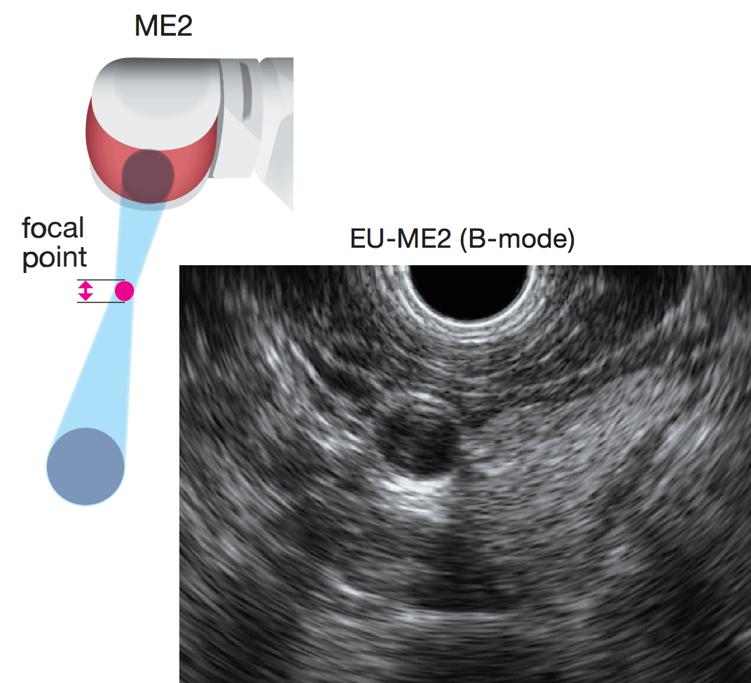
Ultrasound waves are transmitted to a target tissue from the distal end of the ultrasound endoscope, and the EUS image is created based on the returning waves. Sharp focusing beam helps to display minute differences in tissue structure. And thanks to narrower ultrasound wave such as sharp focusing beam, higher quality images can be obtained.
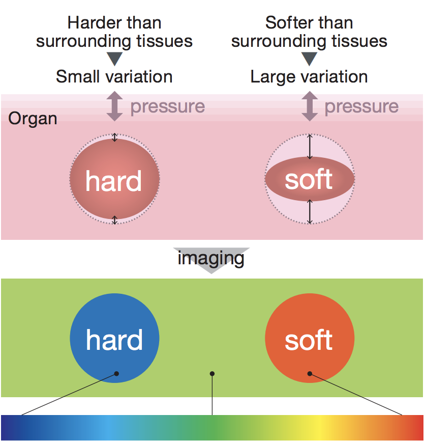
Elastography (ELST)
The main idea of ELST is that stiffness of the tissue gives diagnostic information about the presence or status of the lesion. When pressure is applied to the lesion during the exploration, differences in distortion between hard and soft tissues occur. These differences in distortion are utilized for the real-time analysis of their stiffness. Thus, images obtained by ELST represent tissue elasticity, which may reflect histopathological differences. ELST displays differences in tissue elasticity in the body on a relative scale. This advanced form of ultrasound aims to help the differentiation between benign and malignant changes of tissue and may help to classify tumours.
Tissue Harmonic (THE)
Case 2 Benign leiomyoma
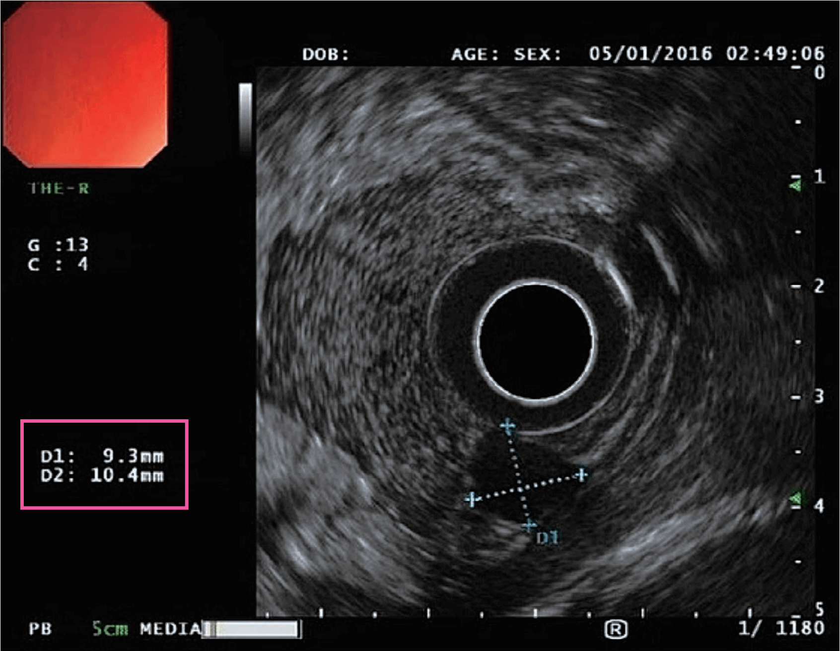
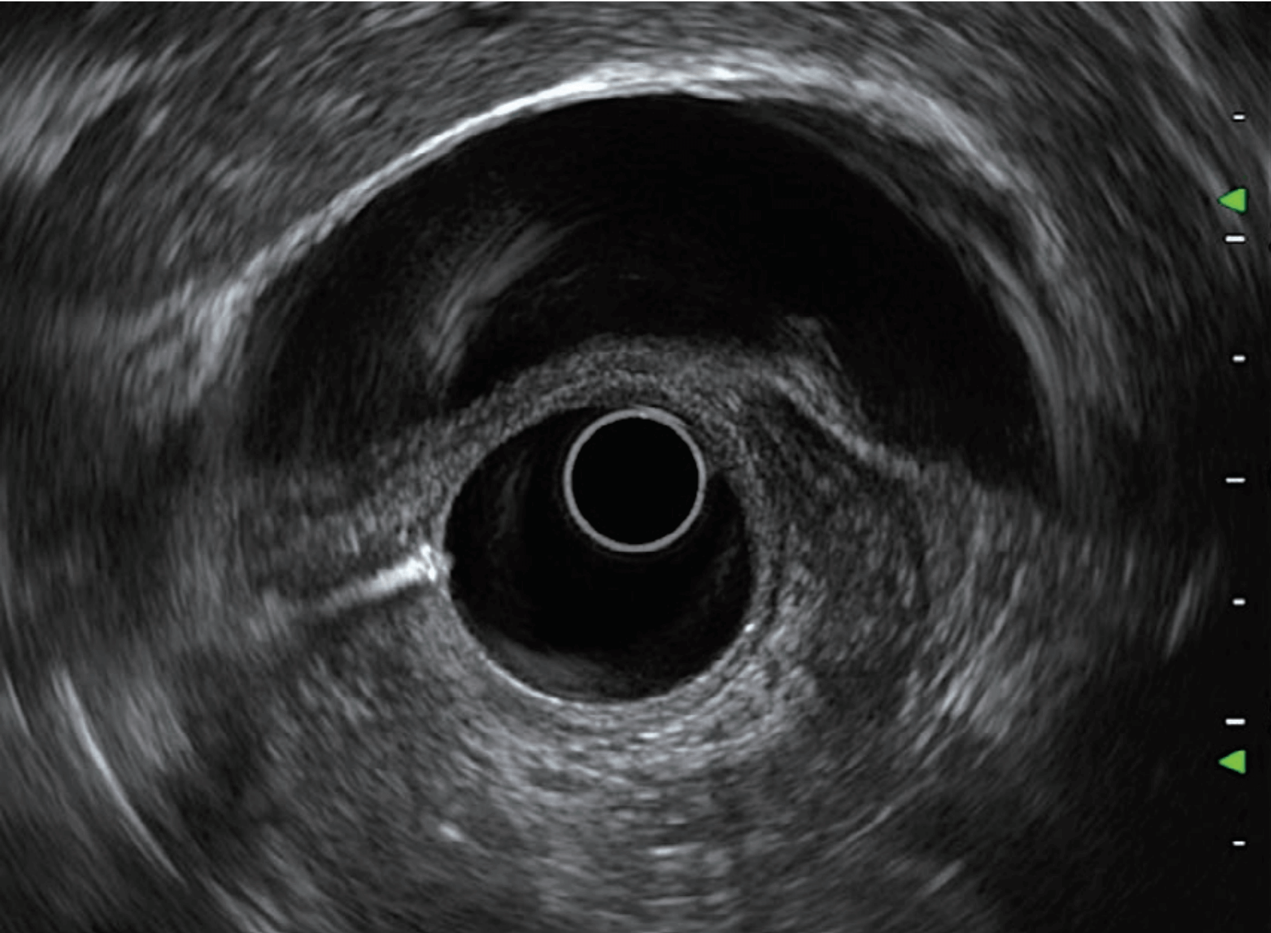
A 43-year-old female patient has hypertension and autoimmune disease. She was diagnosed in 2013 with a gastric leiomyoma < 1 cm. EUS was performed again as part of a surveillance program. THE was used to view the gastrointestinal layers with less artifacts and more contrast. A 9.3 mm x10.4 mm homogenously hypoechoic ovoid lesion with discrete borders was seen from the mucosa to the muscularis propria.
Given that discrete borders were observed and the size of the lesion remained since 2013, the leiomyoma was determined to be benign.
Here, THE proved very useful in producing an ultrasound image with less artifacts. This allowed my team and I to confirm, with greater confidence, that the leiomyoma is still benign.
Technical considerations
Tissue Harmonic (THE)
THE would be helpful to observe a target tissue by reducing the amount of artifacts in the image. THE image is developed by transmitting and receiving signals at different ultrasound frequencies. For example, in THE mode, the EUS scope would transmit ultrasound wave at a certain frequency, but it would be received at a higher frequency. This improves the image quality because body tissue reflects ultrasound waves at a higher frequency than that of what it was initially sent at. This results in clearer images that displays body tissue with less artifacts.
Discussions Future perspectives
ELST and THE are helpful for improving the cyto-pathological yield of pancreatic masses, and following up on patients with benign lesions. As pancreatic masses associated with chronic pancreatitis or incidental pancreatic masses remain diagnostic challenges, ELST and THE may prove useful in such cases. Further studies should be done to demonstrate their differentiating capability.