Laryngeal Cancer (Right Vocal Cord) Aged 71, male
Comment:
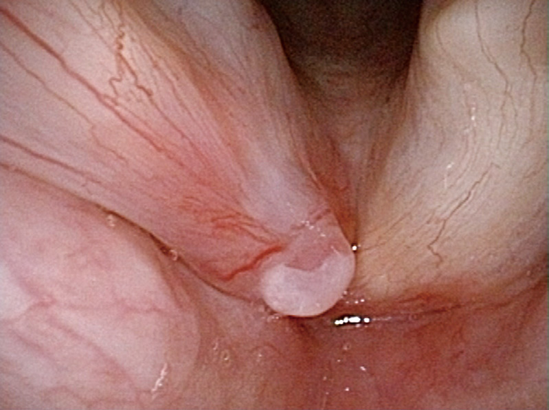
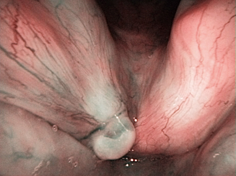
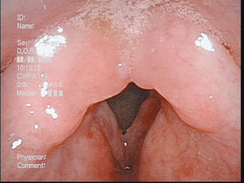
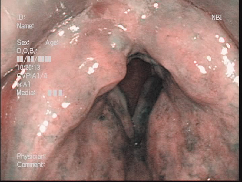
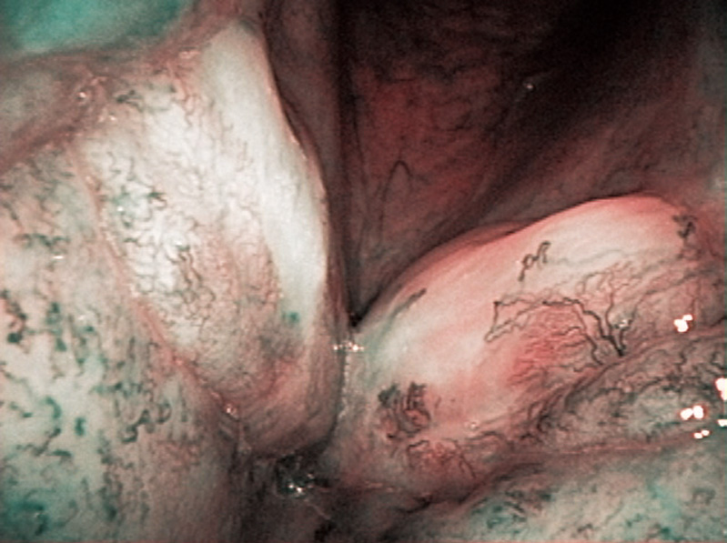
The patient was referred to us complaining hoarseness and a reddening of the right vocal cord. In white light observation, a mild reddish and mucosal irregularities were recognized in the frontal area of the right vocal cord. In the NBI close-focus view, dot-like intraepithelial abnormal vessels localized in the vocal cord were identified.
The lesion was treated with the YAG-laser vaporization under endoscopy (through direct laryngoscopy), and pathologically diagnosed as a squamous cell carcinoma.
After vaporization
Comment:
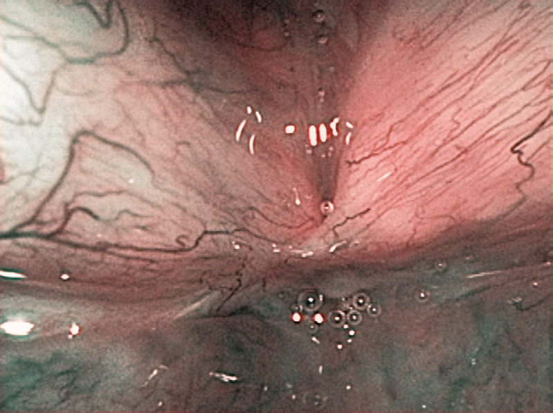
Three months after the laser vaporization, a tumor mass was observed in the same region. However, as no abnormal vascular proliferation was recognized even in the NBI close-focus view, it was diagnosed as a granuloma due to the laser vaporization. Four months after the vaporization, the granulation disappeared and no abnormal vascular proliferation suggesting its recurrence was observed.
Images and comments by Dr. Y. Satou <ENF-VQ>
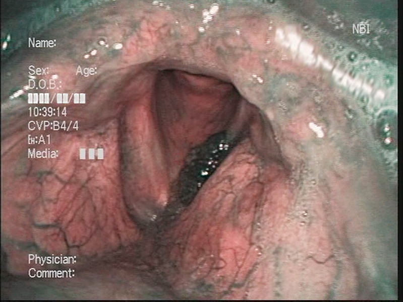
Laryngeal Cancer (Right Vocal Cord) and Vocal Cord Dysplasia (Left Vocal Cord) Aged 67, male
Comment:
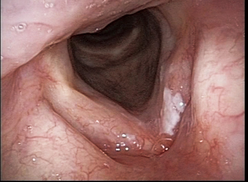
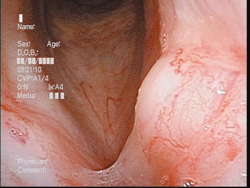
The patient was referred to us by a local hospital with white spots on the left vocal cord. Although the left vocal cord presented obvious leukoplakia, we did not observe any clearly abnormal vessels around the leukoplakia in the NBI close-focus view. On the other hand, and intraepithelial abnormal vessels were identified in the right vocal cord. Therefore a squamous cell carcinoma in the right vocal cord and dysplasia in the left vocal cord were suspected.
general anesthesia
pathology
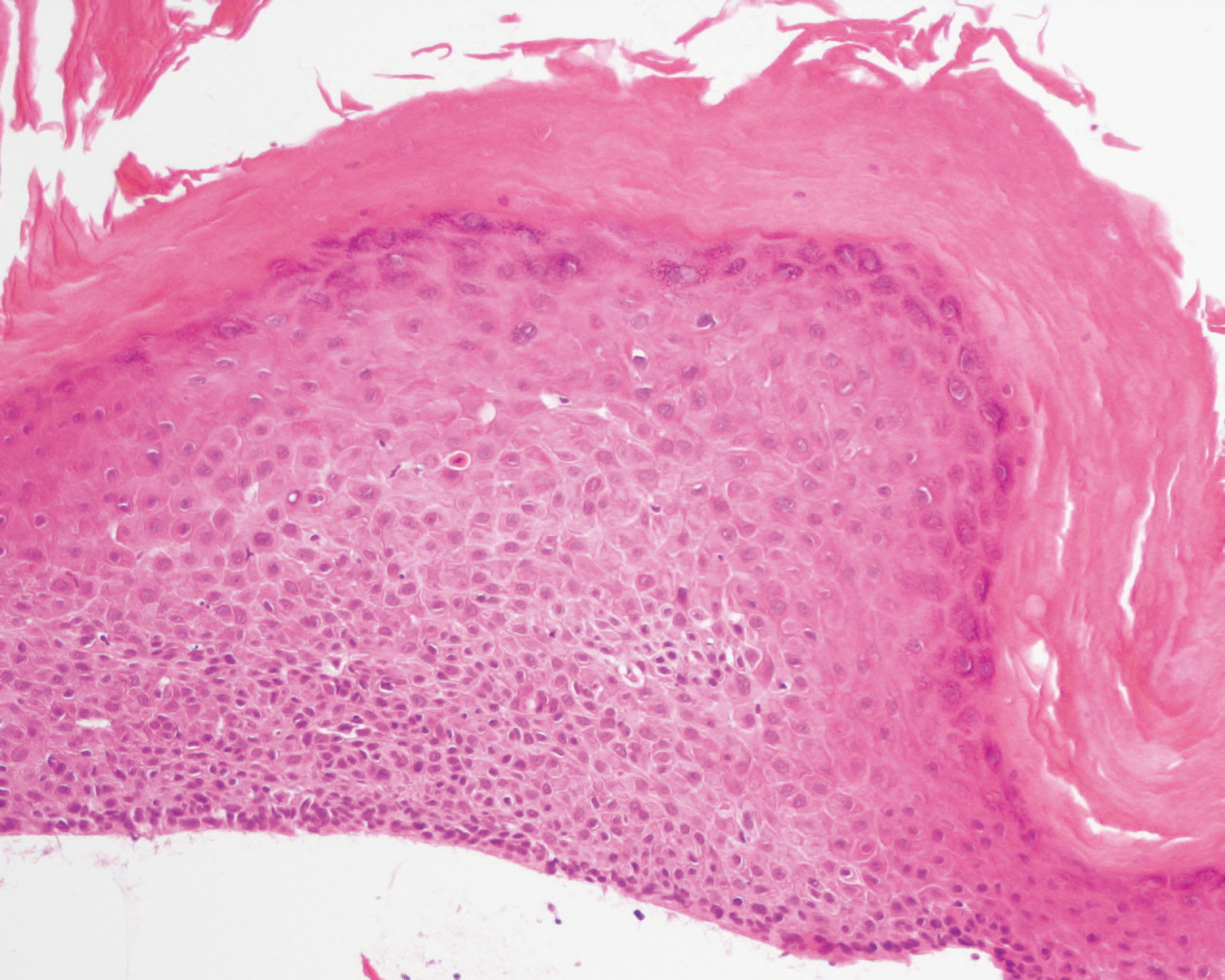
Comment:
These are the peroral endoscopic findings observed under general anesthesia (through the laryngoscope). NBI was useful for the biopsy of the right vocal cord, because it was difficult to recognize the lesion in the white-light view. After the biopsy of the lesions, both vocal cords were vaporized with a YAG laser. The right vocal cord was pathologically diagnosed as an intraepithelial carcinoma and the left vocal cord was a dysplasia (low-grade).
Images and comments by Dr. Y. Satou <ENF-VQ>
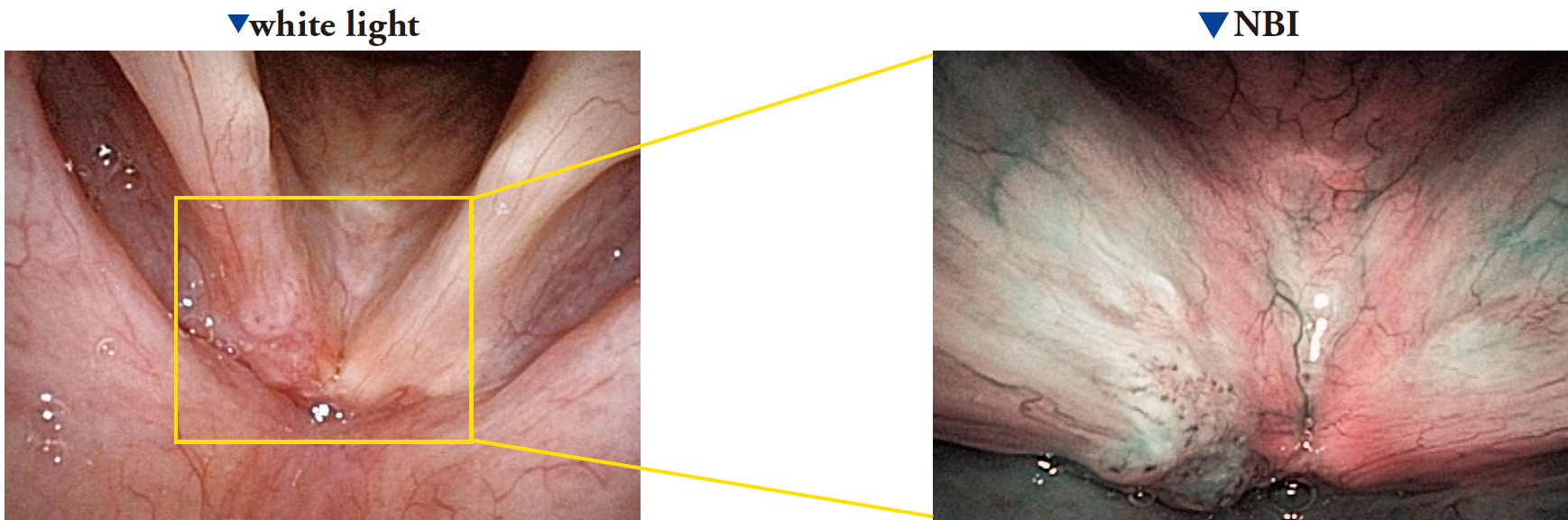
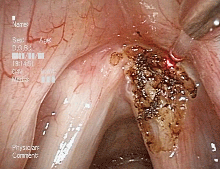
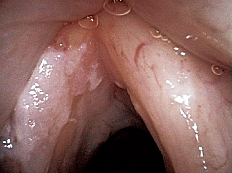
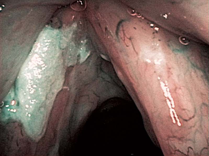
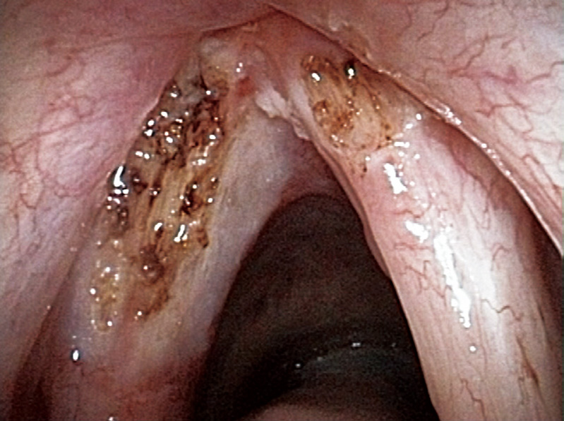
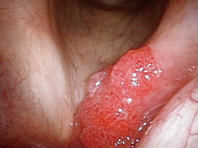
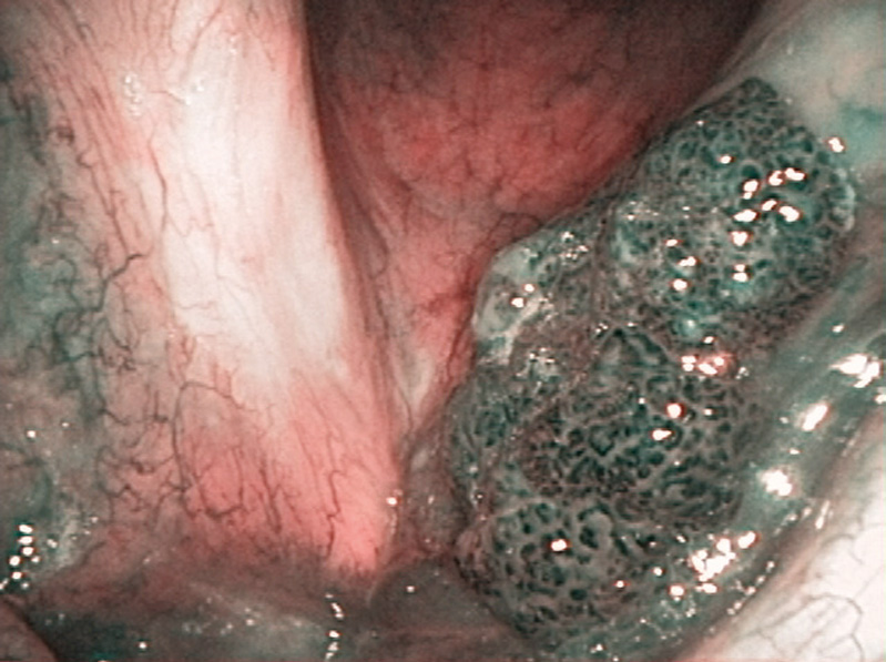
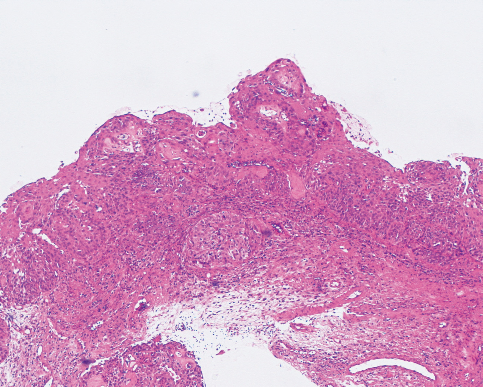
Laryngeal Cancer (Left Vocal Cord) Aged 66, male
close-focus view
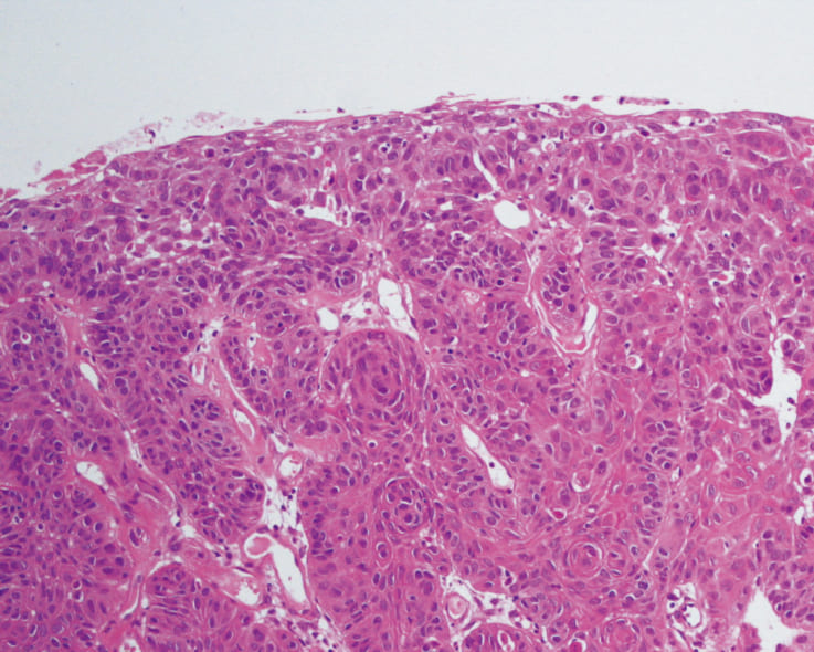
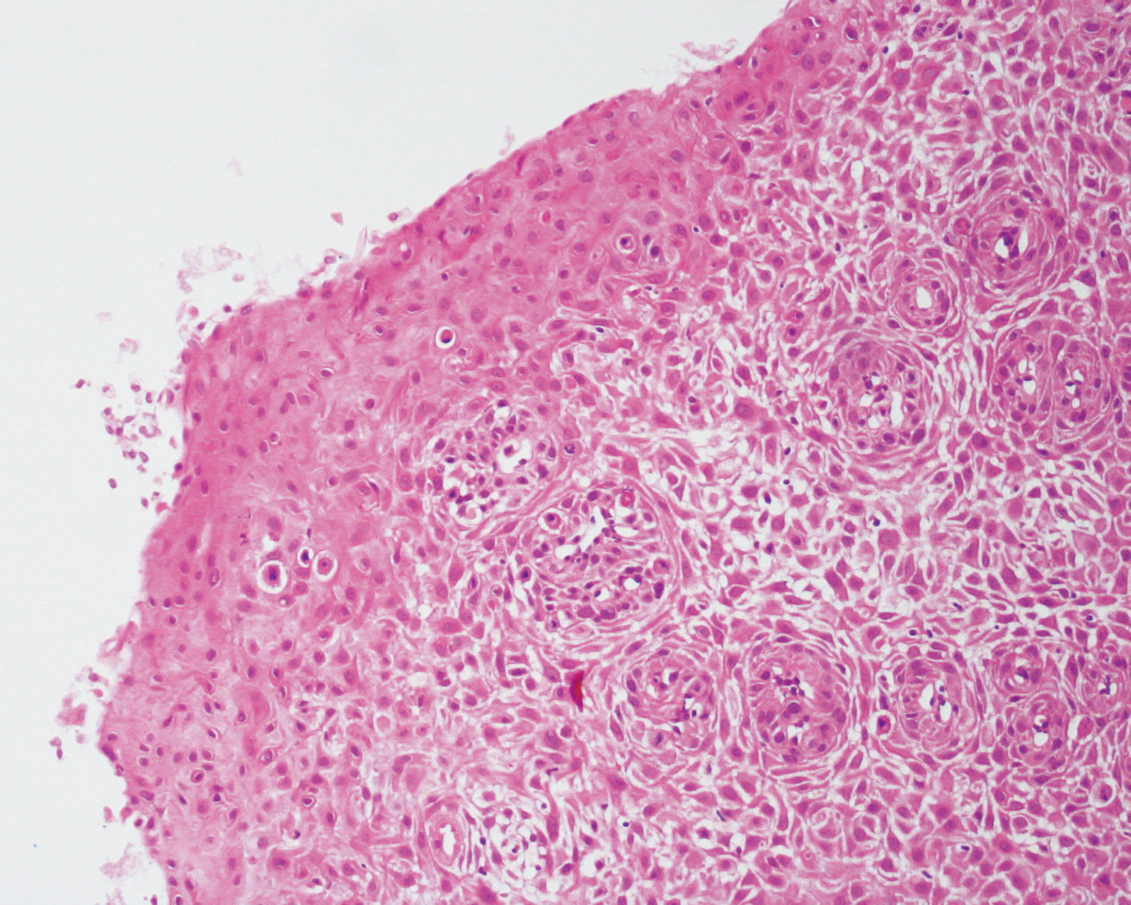
pathology
Comment:
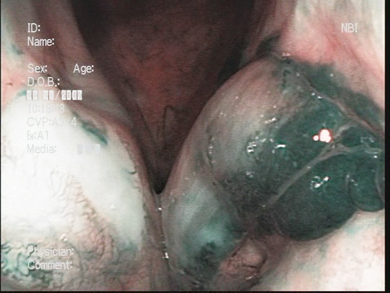
The patient visited a local hospital complaining hoarseness and was then referred to us. A reddish tumor mass was recognized in the left vocal cord and the white light close-focus view identified abnormal vascular proliferation. Weaving and dilatation of abnormal vascular proliferation were more clearly visualized in NBI observation, thereby leading to a diagnosis of a malignant tumor. The lesion was treated with laser depolarization and pathologically diagnosed as a moderately-differentiated squamous cell carcinoma.
Images and comments by Dr. Y. Satou <ENF-VQ>
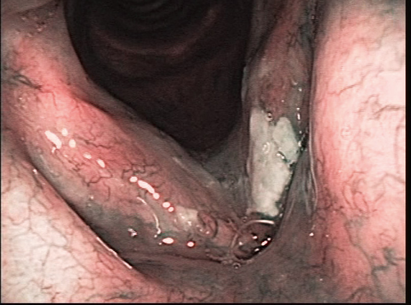
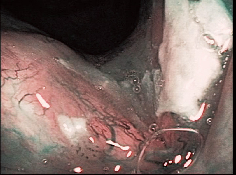
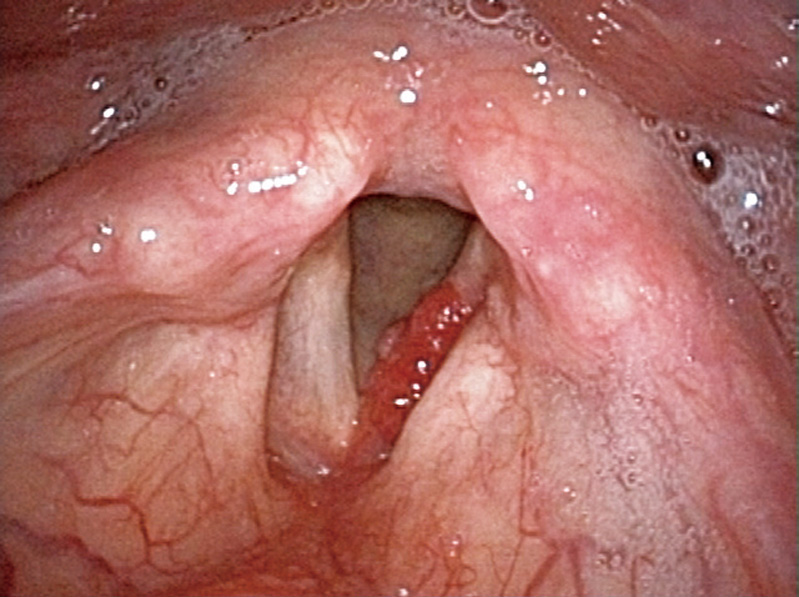
Acute Laryngitis (Left Vocal Cord) Aged 69, male
close-focus view

1 month after vaporization
Comment:
The patient visited us complaining hoarseness after receiving irradiation for a right laryngeal cancer. Reddish swelling was recognized in the left (unaffected side) vocal cord and the entire larynx was edematous due to radiation-induced laryngitis. Since the NBI close-focus view did not show the abnormal vascular proliferation, the lesion was diagnosed as a hematoma caused by acute laryngitis with no malignancy. The reddish lesion disappeared within 1 month as a result of administration of antibiotics and antiphlogistics.
Images and comments by Dr. Y. Satou <ENF-VQ>
- Content Type