Case2 – NSCLC Stage III A (T2N2M0)
Authors:
Prof. Felix JF Herth, MD, PhD, DSc, Thoraxklinik, University of Heidelberg, Germany
Ralf Eberhardt, MD, PhD, Thoraxklinik, University of Heidelberg, Germany
Source:
DVD-ROM ‘Endoscopic Ultrasound – Diagnostics and Staging of Lung Cancer’, Olympus Europa SE & Co. KG, 2013
Patient History
58 years, former smoker (25 cigarettes a day for 25 years) quit smoking 12 years ago.
Persistent cough, suspect of cancer, admitted for diagnosis/staging.
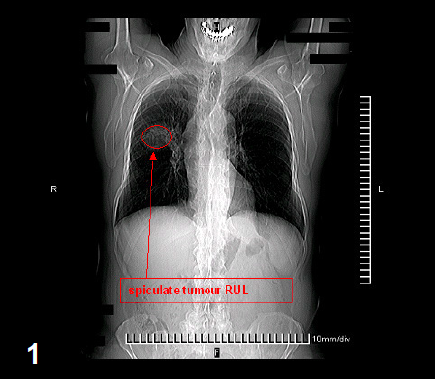
X-ray
Chest X-ray showed 45 mm large spiculate tumor in the right upper lobe [fig. 1].
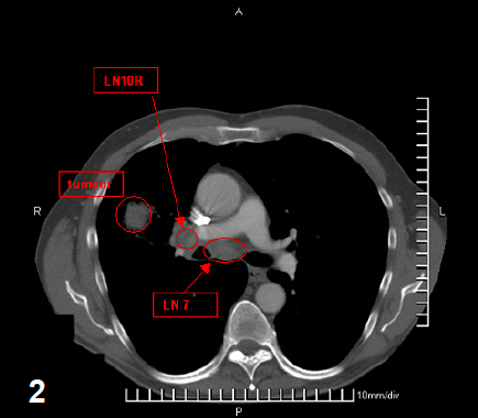
CT
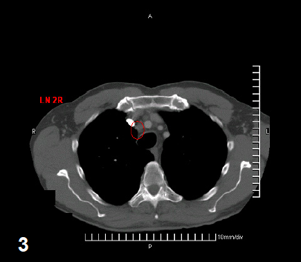
CT showed this tumor and an enlarged hilar (station 10R), subcarinal (station 7) [fig 2] and upper paratracheal lymph node (station 2R) [fig 3].
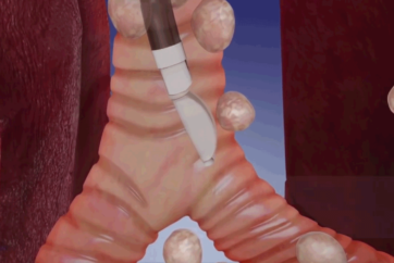
Endobronchial Ultrasound
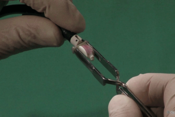
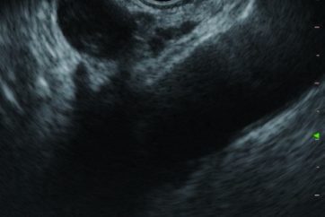
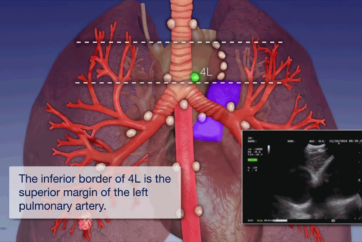
EBUS-TBNA showed enlarged lymph nodes at station 2R [fig 4], 4R, 7 [fig 5], and 10R.
EBUS-TBNA case2-1
2:16
4
EBUS-TBNA case2-2
3:21
5
Cytology
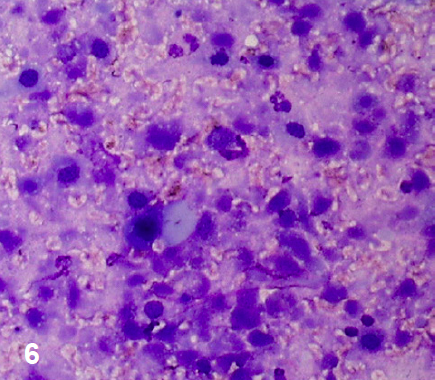
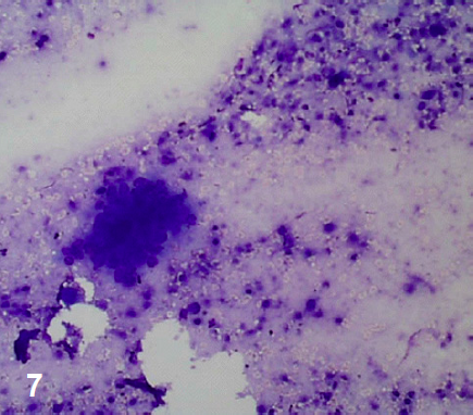
NSCLC (squamous cell carcinoma) [fig. 6,7]
Diagnosis
NSCLC Stage III A (T2N2M0)
Therapy
The patient was referred to chemotherapy.
Benefit
Proper staging and treatment decision;
mediastinoscopy avoided.
- Content Type